Kat Evans, IMV in-house radiographer explains the workings of Digital Radiography (DR)
How DR X-ray Technology Works
All DR X-ray systems comprise of a receptor panel and computer which in the past, were always linked by a cable.
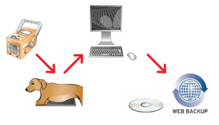
Due to increased portability and speed of image acquisition, DR X-ray technology systems have become a key piece of equipment , particularly for equine vets. Such improvements have enabled vets to provide patients and clients with an increasingly comprehensive service whilst out on the yard.
The increased viability of such technology is opening the way for small animal practices to benefit from the increased efficiencies and time saving advantages.
In addition, installation of such equipment is becoming much simpler since the advent of Automatic Exposure Detection (AED), which means there is no longer any need to wire your generator directly into the DR processor.
Types of DR Technology Available:
Not all DR systems work in the same way. Below is a brief description of each of the 3 main categories:
Charge-coupled devices
There are very few charge-coupled devices found in the veterinary market as the image quality is not great. They are easy to spot as rather than having a thin receptor plate they have quite a ‘deep’ receptor, almost like an old-fashioned TV.
These systems form an image on a large plate, which is then transformed into a digital image, but image quality is often lost during this transfer.
The same technology is used for fluoroscopy systems (also known as C-arms), and the images are ideal if you require a moving image. However sadly they are not as good as so-called ‘flat-panel’ DR or CR.
Direct Digital
Much more common is direct digital radiography, using flat-panel detectors. In the last 5 years the flat panels have become a lot thinner – reducing from about 5cms depth down to only 1.5cm, which has consequently resulted in them becoming lighter and more portable.
There are two types of flat panel:
- Indirect flat panel
- Direct flat panel
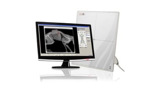
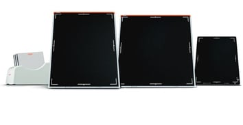
Indirect flat panels are most commonly used and tend to be more sensitive to radiation and with additional advances in technology the image quality resolution has become very high. In addition, most processors will now produce this high-quality image within 5-10 seconds.
Recently, new panels have been developed without any glass involved to improve how rugged they are, but currently these tend to be quite expensive, and are often much heavier.
DR plates are by their nature sensitive to trauma, and when used in equine practice it’s common to put them into a holder to help protect them. In a small animal setting the plate is ideally used in a floating top table, however holders are available where this is not possible.
A DR plate is a highly advanced piece of technology which brings a multitude of benefits to both patient, user and practice. They are built to be used but it is always good to remember to treat them with respect and a little TLC to ensure they will continue serving you for many years to come.
Discover more…
We are dedicated to providing the best equipment, advice, learning, customer care and technical support.
Did you enjoy reading our article? Want to read more about our story and community activities?
Join the discussion. Follow us on Facebook and share your veterinary imaging experience with other vets and industry experts.
Or do you have a particular question regarding any of our products? Our experienced team will be happy to discuss any queries you may have.
Did you enjoy this blog article?