Have you started learning a new skill lately? Many of us will have skills we are learning, improving, or have struggled with for a long time. For some, our interests are fleeting, for others that skill may become a passion, obsession or even a new job.
When beginning the journey to learning something new, we will often ask ourselves: how long will this take? Piano virtuoso or renowned craftsman status was not attained overnight but often takes years of hard work.
When it comes to the skills that we need to perform our role as veterinary surgeons, the rules are the same. Vet school gives us the foundational knowledge, but many skills are improved and perfected over time.
Veterinary ultrasonography is one such skill; despite advances in the technology, it retains an operator-dependant component that requires training and practice. However, many vets can find ultrasound frustrating, and may not use the equipment to its full potential. We may blame ourselves rather than recognise that time and experience are needed to improve. In human medicine, the Royal College of Radiologists’ “Ultrasound training recommendations for medical and surgical specialities” sets out proposals for minimum training requirements. The training requirements for level 1, wherein common examinations can be performed safely and accurately, often involve a minimum recommendation of 250 scans. It is worth noting that these recommendations are based on body system specific examinations and the recommendations for higher levels may involve practitioners performing an additional 600+ scans as a minimum.
So, how can we help ourselves to improve?
Competency at ultrasound can be divided into four broad components:
- Identifying when to perform an ultrasound examination
- Technical skill in image acquisition (factors reported in studies assessing errors in human ultrasonography include; lack of knowledge of the technical equipment, use of inappropriate transducers and inadequate optimisation of the images1)
- Interpreting the images acquired
- Incorporating findings into clinical decision making
Each of the categories can be improved by a variety of methods:
- Get familiar with your machine.
Getting started with ultrasonography can be daunting, especially as most modern systems have a plethora of modes and functions available.
However, the majority of systems share a core set of controls that are used to optimise the image quality during an examination. These are the gain, time gain compensation, frequency, depth, and focal zone controls.
Becoming familiar with the location and function of each will help to enhance the quality of your images.
Similarly, understanding the relationship between your ultrasound transducer and the on-screen image is key. Take time to get to know which transducers are available, the pros/cons of each and the location of any transducer markers – how does that marker relate to the on-screen view?
Also, remember that most modern systems have pre-sets ready for certain examination types. Use these, as they will give you a good starting point for most examinations.
2. Prep properly.
Although time is always against us, some of the most common image quality problems are caused by poor patient preparation. Ensuring that patients are adequately clipped(Figure 1.)and cleaned, and that the gel has had sufficient time to soak in can improve image quality and make our examinationsmore rewarding.Remember that analgesia and chemical restraint can aid patient comfort and will give us the time we need to complete a thorough examination.
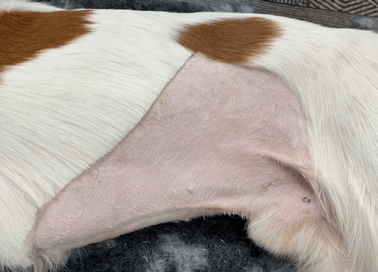
Figure 1. Make sure to clip patients adequately. The image above shows the standard clip for performing abdominal ultrasound on a small to medium sized dog. The same area would be clipped on the right abdomen as well.
3. Be consistent
Regardless of the examination we are performing, approaching each patient in a systematic, routine manner can help. Consider creating or using a checklist for your examinations; by doing so it can help us approach each examination in a methodical way (Figure 2.).
Sticking to a prescribed method of assessing each area or organ in turn can help us to understand what normal looks like and how parts of the anatomy are positioned.
Likewise, try to keep the orientation of the transducer marker consistent. This way, we can quickly appreciate how our transducer movements will affect our images.
Over time, consistency in our approaches starts to make examinations faster and more natural. We also develop a more unconscious understanding of the relationship between the two-dimensional ultrasound image and the three-dimensional anatomy.
Consistency is also important when interpreting images. Where changes are present, we can assess them using a common set of factors like the Röntgen signs used in radiography:
- Size
- Shape
- Number
- Location
- Margination
- Echogenicity
Using these terms to define changes can help with the formation of differential lists and with the interpretation of images using reference material.
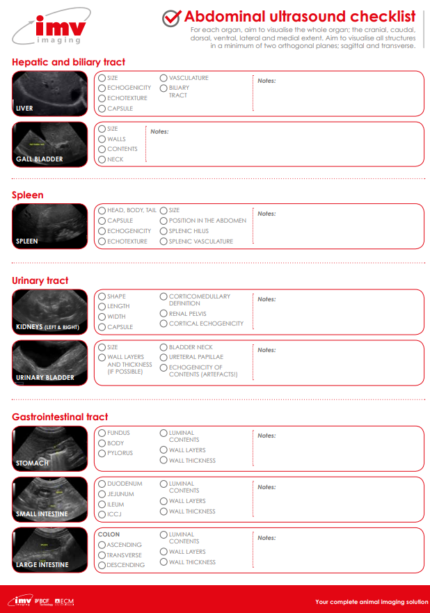
Figure 2. A checklist is a great way of keeping examinations on track and being methodical. This one can be downloaded from the IMV imaging website.
4. Practice makes perfect
Once we are familiar with our machine and have a routine approach to patients, nothing will help more than performing examinations. Do you have a pet who might let you practice? Or could you borrow one from friends or family? There may even be patients coming in for routine examinations or procedures that could be safely examined.
There are also many recipes available for ultrasound phantoms. These are especially useful for practicing ultrasound-guided needle sampling and can be made from easily acquired household ingredients.
5. Learn from the best
Many institutions and CPD providers offer practical ultrasound courses. The knowledge gained from these can help form the routines and approaches that create a good foundation for starting out in ultrasonography. Alternatively, the courses may offer the opportunity to learn more advanced techniques, taking examinations to the next level.
Similarly, there are many fantastic reference texts available. These can give examples of the common approaches and appearance of normal anatomy as well as pathological conditions. Having a good textbook can be very helpful when we come to interpret our images.
If possible, consider work experience with practitioners working at referral centres. Although they may have access to high end equipment, seeing specialist’s perform scans can help us develop our own approaches and can highlight the different applications of ultrasound.
6. Save your images
Saving images or videos from patients is an important part of clinical record keeping (Figure 3.). Yet it can also help us develop our image interpretation skills; we can review the images later (possibly when we have a little more time) and this may allow us to appreciate details that we could have missed. It also allows us to compare our images to those in reference materials. Likewise, we can save any measurements taken, as well as labelling anatomy for reference.
Videos (cine-loops) are especially useful. These may offer a more recognisable view of the area of interest and aid interpretation in cases where images need to be passed onto specialists for advice.
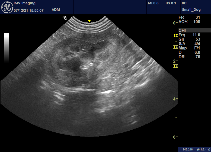
Figure 3. This image shows the left kidney in a dog. By saving our images we can keep records of our examination findings and refer back to them later.
7. Join a community
There are online communities that focus on many different areas. Veterinary imaging is no different and these groups can give access to other like-minded professionals for case discussions and advice, as well as offering resources including webinars and journal clubs.
8. Go big
If diagnostic imaging becomes more of a passion, then consider the various post graduate certificates available. Similarly, many institutions offer internships which may lead to further qualification and specialisation opportunities.
The information listed above offers a section of ways to improve our ultrasound skills and is not exhaustive. However, they offer a good starting point for future improvement. Remember, like any skill, our ability to perform ultrasound will improve with practice and time, and with both, our examinations will become more rewarding.
References
- Pinto A., Pinto F., Faggian A., Rubini G., Caranci F., Macarini L., Genovese E.A., Brunese L. (2013) Sources of error in emergency ultrasonography. Critical Ultrasound Journal. 5 (Suppl 1).