Monday, April 1, 2019
Indications
- Investigation of pain causing lameness localised to the fetlock region by clinical signs and/or diagnostic analgesia.
- Investigation of traumatic/penetrating wounds to the fetlock region.
- May be requested as part of a pre-purchase examination.
- Assessment of angular limb deformities.
- Monitoring and re-evaluation of pathology.
Patient preparation
- Ensure the hair coat is clean as debris will appear radiopaque on radiographs and may mimic or mask abnormalities.
- Radiography of this region is usually well-tolerated. However, sedation will usually aid the acquisition of well positioned and good quality radiographs and reduce procedure time, number of repeated acquisitions and risk to personnel and equipment.
- The horse should be weight-bearing evenly on the forelimbs with the metacarpus of the limb to be radiographed as vertical as possible in both planes (i.e. when viewed from both the front and the side). The same principles of positioning apply when radiographing the hindlimb fetlock.
Radiographic views
There are four standard views of the fetlock joint:
- Lateromedial
- Dorsopalmar(/plantar)
- Dorsolateral-palmaro(/plantaro)medial oblique
- Dorsomedial-palmaro(/plantaro)lateral oblique
There are various other additional views for identifying specific lesions or visualising specific areas.
The technique for radiography of metacarpophalangeal joint (forelimb) is described below, however the same principles apply for radiography of the metatarsophalangeal joint (hindlimb).
Lateromedial (LM)
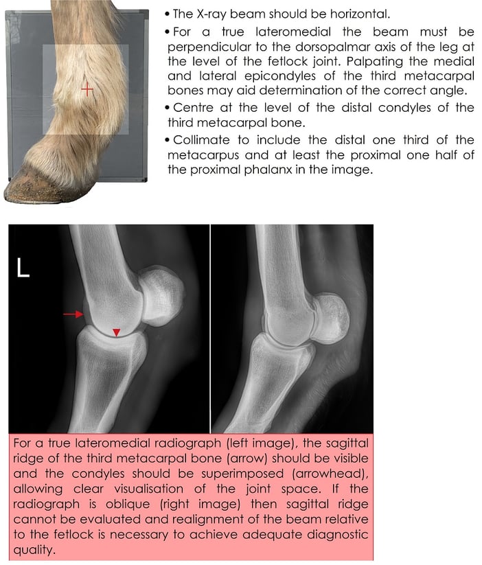
Flexed lateromedial

Dorsopalmar (DP)

Dorsolateral-palmaromedial oblique (DLPMO)

Dorsomedial-palmarolateral oblique (DMPLO)

Additional radiographic views
Angled-down obliques (DPrM-PaDiLO and DPrL-PaDiMO)
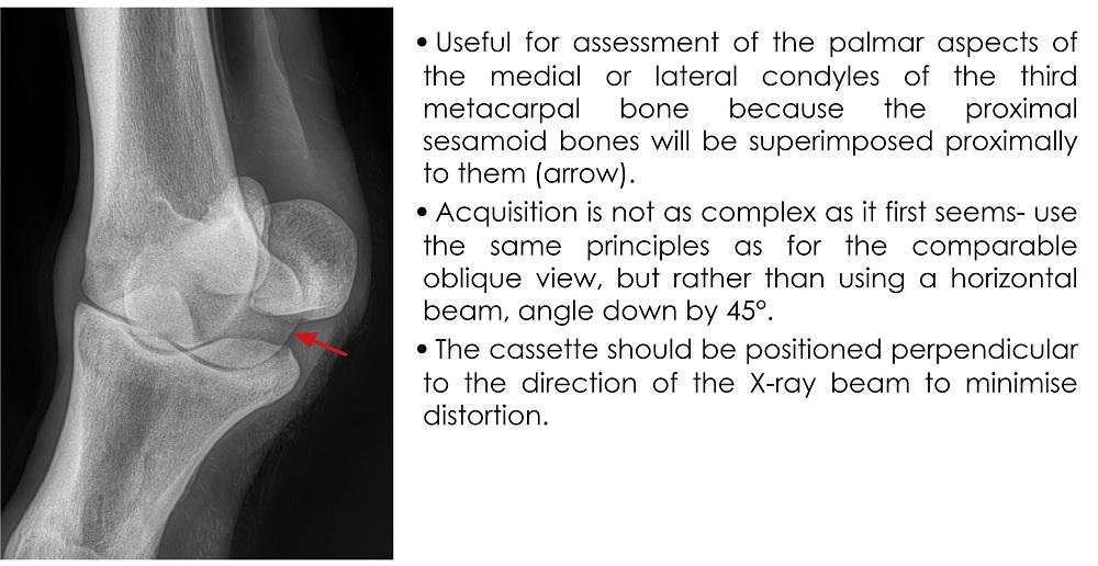
Flexed dorsopalmar

Skyline of the metacarpophalangeal joint

We hope you enjoyed this blog post!


Don't forget to share! 👇
IMV imaging Service Contracts… More than just an annual service
Improving elbow radiography - the importance of positioning and exposure