Ultrasound Frequency Range
With regard to image quality related to frequency, there is a trade-off between resolution and penetration.
A higher frequency produces a higher resolution image, but the ultrasound wave will not penetrate as deeply into the patient. A lower frequency means that you will get a decrease in image resolution, but the ultrasound beam will be able to penetrate to a greater distance into the patient. It is important to strike a balance between these two factors to achieve an acceptable image of the area that you want to evaluate.
Most small animal ultrasound scans are performed at a depth of 2 – 15 centimetres. A frequency range of 4 – 10 MHz should allow you to scan within these depths.
A modern electronic probe can scan at multiple frequencies while an older mechanical probe often only operates at a single, fixed frequency.
Types of veterinary ultrasound probes
Micro-convex veterinary ultrasound probe
This is the main probe that is used for small animal abdominal scanning. A smaller footprint allows better surface contact to be achieved which will result in better image quality. Compared to using a large convex probe, it is easier to scan through rib spaces and underneath the costal arch with this probe and the frequency range is always of a higher frequency.
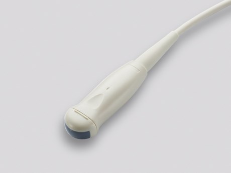
GE Mindray M5 6C2s micro convex probe
Convex veterinary ultrasound probe
A convex probe has a large point of contact with the skin surface, with the ultrasound beam spreading to a wider field of view within the patient, giving a sector-shaped image. This type of probe is often supplied with ex-NHS systems as it is good for human abdomens and is always used in maternity departments. Maternity units have the highest turnover of systems so are commonly available second hand along with their probes. However, these probes are really not suitable for small animal work as they are too large to fit into areas like the intercostal spaces for viewing the heart or lateral lobes of the liver, xiphisternum for scanning the liver and behind the costal arch for viewing the kidneys. They also typically have a lower frequency range (3.0-5.0 MHz), therefore the resolution you can achieve will be less than with other types of probe. Although they can be useful for scanning the liver in extremely large dogs!
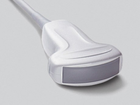
Sonosite M-turbo C60 radius convex probe
Linear veterinary ultrasound probe
Linear probes are primarily used for superficial abdominal structures, especially in cats, such as the small intestine and the kidney. It is also excellent for imaging the eyes! Linear probes normally give a higher resolution image than convex probes for several reasons:
- They often scan at higher frequencies
- The ultrasound beams do not diverge as they travel away from the probe. They travel in a straight line, parallel to the adjacent beams.
One drawback is that they often have quite large footprints, so for small animals and exotics a hockey stick linear probe is preferable (see below).
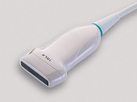
Mindray M5 10L4s linear probe
Hockey Stick veterinary ultrasound probe
Hockey Stick probes are linear probes with an ultra-small footprint. They operate at a higher frequency range, but the penetration ability is poor. They are designed as an intra-operative probe, meaning that it would be used directly on an organ during surgery. These features make it a superb choice for scanning exotic species, musculoskeletal scanning in small animals (such as the biceps tendon) and for ophthalmology.
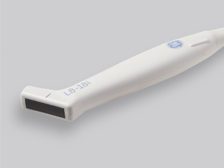
GE Logiq S7 expert L8 18i linear hockey stick probe
Phased Array veterinary ultrasound probe
A Phased Array probe would be the probe of choice for echocardiography.
The ultrasound beam comes from a smaller point of origin, making it easier to pass between the rib- spaces of the patient. The probe can generate a very high pulse repetition, which allows the scanner to produce a scan with an extremely high frame rate. The benefit of this is that when scanning a rapidly moving heart, the image can be played back frame by frame and small changes in position can be observed and detected.
Phased array probes can also perform better Doppler scans.
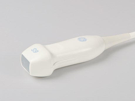
GE Vivid q 6S RS array probe
Matrix veterinary ultrasound probes
Matrix Probes are a relatively new addition to ultrasound scanning. The crystals in the probe are arranged in several linear arrays forming a matrix. This improves lateral and axial resolution and allows focus in two dimensions rather than just one. In other words, these probes produce a very high quality, high resolution image!
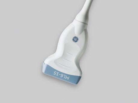
GE Logiq S7 ML6 linear matrix probe
Final thoughts and advice
The most important thing to remember is to not drop the probe! They house the peizoelectric crystals that produce the ultrasound waves. Although these not only break easily, they are almost impossible to repair. The probe is also normally the most expensive part of a scanner, with the price of a probe ranging from £1,200 to more than £5,000.
Keeping the probes clean and making sure to follow the cleaning guidelines in the product manual (with a list of approved cleaning agents) is also important. Probes do not like spirits, so restrict the use of spirit to biopsies and FNAs. Prolonged exposure to spirits can cause the surface of the probe to dry out, harden and crack/peel which will affect the image quality. We recommend thinking of the surface of the probe as a camera lens!
I would also encourage people not to remove the probe from the machine unless the machine is switched off. If this happens, you may get a surge of power through the machine which can cause problems if the probe is not attached. Some machines are safe to change probes whilst on, but the image should almost always be frozen before detaching a probe. Another thing to remember is that probes are generally not interchangeable between different scanners, although there are some exceptions to this rule between certain scanners made by the same manufacturer.
I would recommend…
Always try out anything before committing to buy to make sure it suits your needs!
Discover more…
We are dedicated to providing the best equipment, advice, learning, customer care and technical support.
Did you enjoy reading our article? Want to read more about our story and community activities?
Join the discussion. Follow us on Facebook and share your veterinary imaging experience with other vets and industry experts.
Or do you have a particular question regarding any of our products? Our experienced team will be happy to discuss any queries you may have.